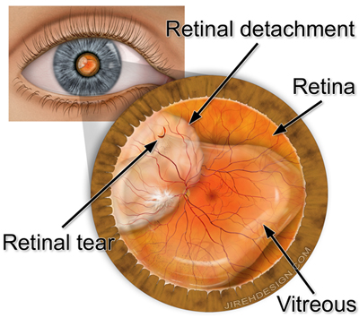
A macular hole is a condition that affects the center of the retina, called the macula. The macula is responsible for central vision, which is crucial for activities such as reading, driving, and recognizing fine details. A macular hole is characterized by a small break or opening in the macula, leading to visual distortion and potential loss of central vision.
Key points about macular holes:
- Formation: Macular holes typically develop due to the aging process and changes in the vitreous gel within the eye. The vitreous gel can pull away from the retina as a person gets older. In some cases, this separation can create tension on the macula, leading to the formation of a hole.
- Symptoms: The early stages of a macular hole might not cause noticeable symptoms, but as the hole progresses, individuals may experience the following visual changes:
- Blurred or distorted central vision
- A dark or empty spot in the center of the vision (like looking through a hole)
- Difficulty with tasks that require detailed vision, such as reading or recognizing faces
- Types: Macular holes are classified into different stages based on their size and severity:
- Stage 1: Foveal detachment (incomplete hole)
- Stage 2: Partial-thickness hole
- Stage 3: Full-thickness hole (with a diameter less than or equal to 400 micrometers)
- Stage 4: Full-thickness hole (with a diameter greater than 400 micrometers)
- Diagnosis: An eye doctor (ophthalmologist) can diagnose a macular hole through a comprehensive eye examination, including dilated pupil examination and imaging tests such as optical coherence tomography (OCT) to visualize the layers of the retina.
- Treatment: The primary treatment for a macular hole is surgery. The goal of surgery is to close the hole and restore as much central vision as possible. Common surgical procedures include:
- Vitrectomy: This procedure involves removing the vitreous gel and any traction it exerts on the macula. The surgeon may also use gas or silicone oil to hold the retina in place while it heals.
- Membrane Peel: During a vitrectomy, the surgeon might also peel away any scar tissue or membranes that have formed on the surface of the retina.
- Recovery: Recovery from macular hole surgery can take several weeks to months. Visual improvement may not be immediate, and final visual outcomes can vary based on the size and severity of the hole and the success of the surgery.
Early detection and intervention are important for better outcomes in treating macular holes. Regular eye exams, especially for individuals at higher risk, such as those with certain medical conditions or a family history of eye disorders, can aid in early diagnosis and appropriate management. If you notice any changes in your vision, especially in your central vision, it’s important to consult an eye care professional for evaluation and guidance.
It is advisable to discuss your eye related queries with an eye care professional Dr. Vaidya, Eye Specialist in Andheri at Dr. Vaidya Eye Hospital for appropriate monitoring and guidance.


