The diagnosis of varicose veins typically involves a combination of a physical examination, medical history review, and, in some cases, additional diagnostic tests. Here is an overview of the diagnostic process for varicose veins:
- Medical History: The healthcare provider will inquire about your medical history, including any symptoms you may be experiencing, family history of varicose veins, and any factors that may contribute to vein issues.
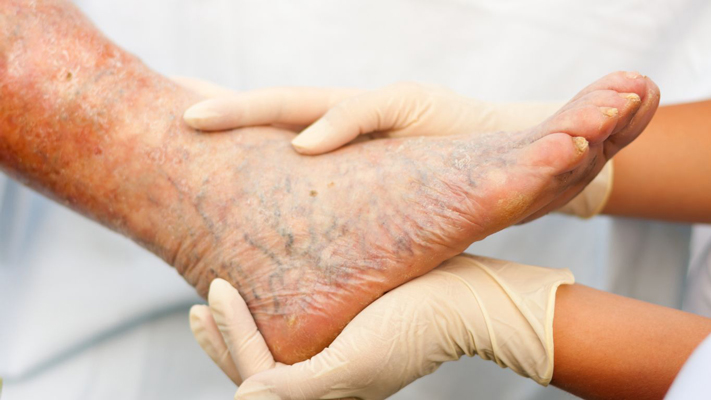
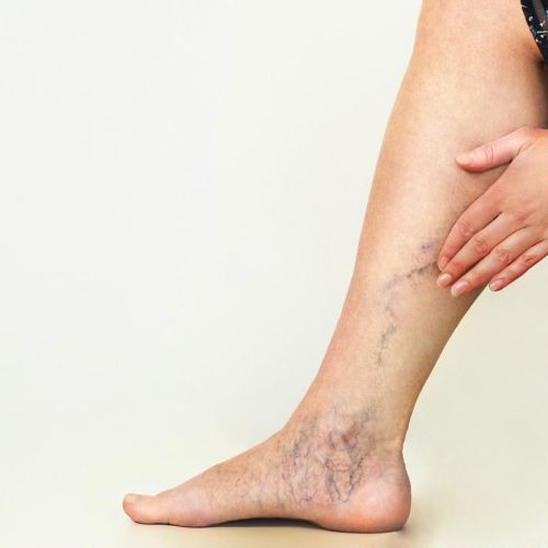
- Physical Examination: A thorough physical examination is conducted to assess the appearance of the veins and identify any signs of varicose veins. The examination may include inspecting the legs while standing and sitting and checking for swelling, skin changes, or ulcers.
- Symptom Assessment: The healthcare provider will ask about specific symptoms such as pain, aching, heaviness, cramping, or swelling in the legs. The severity and duration of symptoms are considered in the diagnostic process.
- Risk Factors Assessment: Identification of risk factors, such as age, gender, family history, pregnancy, obesity, and occupation (long periods of standing or sitting), helps in understanding the potential causes of varicose veins.
- Ultrasound Imaging: Doppler ultrasound is a common diagnostic test for varicose veins. This non-invasive imaging technique uses sound waves to create images of the blood flow in the veins. It helps assess the structure and function of the veins and identify any valve dysfunction or blood flow abnormalities.
- Venous Reflux Testing: Venous reflux testing, often performed as part of the ultrasound examination, assesses the backward flow of blood in the veins. This helps identify reflux, a common factor in varicose veins.
- Photoplethysmography (PPG): PPG is a test that measures changes in blood volume in the legs. It is sometimes used to assess venous insufficiency and blood flow abnormalities.
- CT or MRI Scan (in certain cases): While less common, computed tomography (CT) or magnetic resonance imaging (MRI) scans may be used in specific cases to provide detailed images of the veins and surrounding structures.
- Contrast Venography (rarely used): Contrast venography involves injecting a contrast dye into the veins, followed by X-ray imaging. This invasive test is rarely used but may be considered in complex cases.
The combination of the medical history, physical examination, and imaging studies allows the healthcare provider to make an accurate diagnosis, assess the severity of the condition, and develop an appropriate treatment plan. It’s essential for individuals experiencing symptoms or concerns related to varicose veins to seek medical attention for a thorough evaluation by a healthcare professional specializing in vascular conditions.
Discover cutting-edge interventional radiology services with Dr. Kunal Arora, renowned for his expertise and commitment to exceptional patient care. As the leading Interventional Radiologist in Mumbai, Dr. Arora specializes in providing advanced, minimally invasive treatments for various vascular conditions.


